Overview
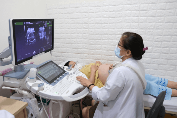
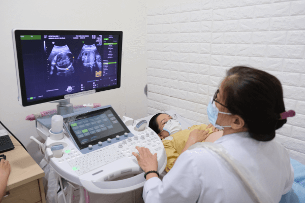
Thien Nhan owns the ultra-premium Voluson Expert 22 4D Obstetric Ultrasound System. This is the most modern obstetric and gynecological ultrasound model from GE – USA, and the first in the Central and Central Highlands region.
The Voluson Expert 22 Ultra-Premium 4D OB Ultrasound at Thien Nhan Hospital
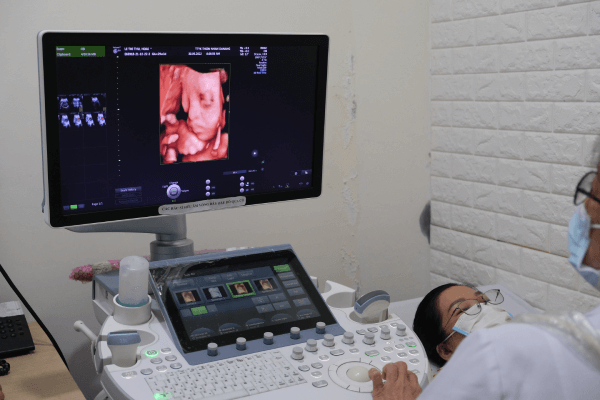
- Sharp, vivid fetal images enable the detection of abnormalities in the smallest details during early pregnancy via UltraHD technology.
- Accurate diagnosis of fetal congenital abnormalities, especially heart defects and central nervous system anomalies, using MRI-like imaging and HDlive, HD Flow Doppler.
- Visual and precise detection of fetal vascular abnormalities (e.g., fetal cardiac defects, true cord knots, placenta accreta/percreta) using Stic Doppler technique.
- The breakthrough Augment technology assists in examining overweight clients, those with previous surgical scars, or surface structures that impede sound wave signals.
- Early classification of multiple pregnancies and detection of placental abnormalities using HDlive Silhouette on the vaginal probe.
- Accurate diagnosis and differentiation of congenital uterine/ovarian anomalies, abnormal first-trimester pregnancies, and ovarian masses using the 3D vaginal probe.
Superior Value of the 4D Voluson Expert 22
In Fetal Ultrasound
- Detection of birth defects starting from the first trimester.
- Accurate detection and diagnosis of fetal morphological abnormalities across all stages: In-depth evaluation of fetal cardiac defects and the central nervous system.
- 3D/4D imaging allows for 3D Print products, bringing joy to parents.

In Gynecology, especially IVF
- Accurate diagnosis of congenital uterine anomalies, such as uterine didelphys or septate uterus.
- Accurate diagnosis of uterine cavity pathologies like polyps or intrauterine adhesions, and precise placement evaluation of IUDs.
- Notably, diagnosis of hydrosalpinx using 3D sonohysterography with a contrast agent.