Overview
Since its establishment, Thien Nhan has aimed to bring the most advanced medical techniques closer to the people of Da Nang and Central Vietnam. Therefore, from the very beginning, Thien Nhan selected top-tier medical equipment to keep pace with global medical development. The 3.0 Tesla MRI system at Thien Nhan Da Nang is a testament to Thien Nhan Hospital’s pioneering step in technology.
Magnetic Resonance Imaging (MRI)
Magnetic Resonance Imaging (MRI) is a technique that uses a strong magnetic field and radio waves to create detailed images inside the body. It is currently one of the most modern techniques for detecting tumors, metastases, inflammation, and other pathological tissue changes at an early stage.
MRI Scanning at Thien Nhan Hospital

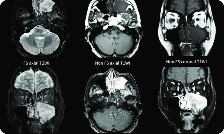
Head and Neck MRI
Scanning of the brain, neck region, blood vessels, cervical spine, temporomandibular joint, and ear-throat area. High image quality, low artifact noise.
Advanced applications for Brain MRI:
- Perfusion MRI
- Functional MRI
- Inner Ear MRI
- Spectroscopy MRI – Diffusion Tensor Imaging (DTI)
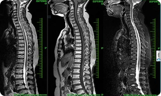
Spine MRI
- Ultra-high resolution imaging of the cervical, thoracic, and lumbar spine
- Reduces artifacts caused by metallic implants
Advanced Applications:
- Automatic whole-spine curvature reconstruction
- Nerve plexus imaging
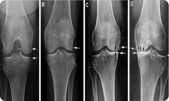
Musculoskeletal MRI
- Specialized 18-channel H3 coil, the most modern in Central Vietnam
- Imaging of the smallest parts to the largest joints with the highest image quality
- 3D knee joint imaging, reducing artifacts caused by metallic implants
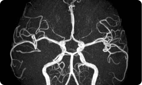
Vascular MRI
- 36-channel peripheral and full lower extremity vascular coil
- High accuracy
- Fast, clear imaging of intracranial/extracranial, visceral, and peripheral vessels for accurate diagnosis of vascular stenosis, occlusion, and malformations without contrast medium
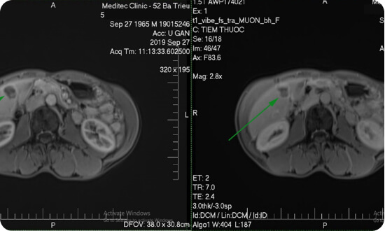
Abdominal MRI
- Reduces respiratory motion artifacts, displaying lesions with high resolution
- Reduces noise and ensures signal intensity homogeneity for accurate diagnostic efficiency
- Contrast injection: Automatic injector, CareBolus software precisely controls post-injection timing, creating dynamic ce-MRA images for 3D visualization over time
- Quantification of liver iron concentration (Advanced)
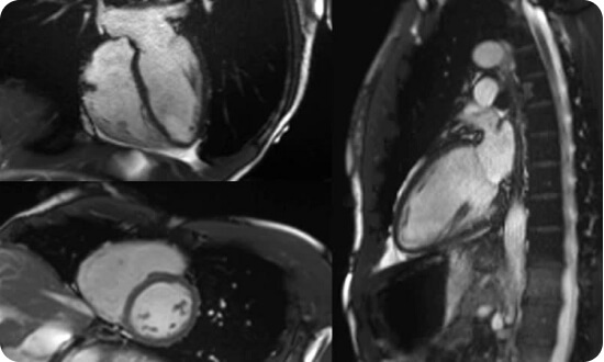
Cardiac MRI
- Breath-hold and free-breathing control to enhance contrast between structures and vessels
- Dynamic imaging allows high-resolution cardiac valve assessment
- High-quality tissue characterization
- High-resolution stress and rest cardiac protocols
- Excellent assessment of myocardial diseases, valvular heart disease, etc.
- Pre-surgical evaluation for congenital heart diseases
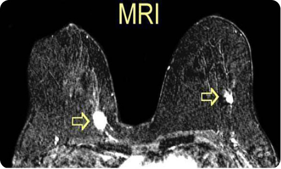
Breast MRI
- High-resolution 2D and 3D sequences
- Sequences for silicone implant evaluation, including perinipple reconstruction software
- Tumor Spectroscopy MRI
- Most accurate assessment of breast lesions with the highest resolution
Whole-Body MRI
- Screening for tumors and other diseases from head to toe: brain, neck region, spine, liver, biliary system, pancreas, uterus/adnexa, prostate, etc.
Screening and TNM staging assessment
- Assessment of local invasion and distant metastasis, comparable to PET/CT imaging
- Helps doctors establish appropriate treatment protocols
- Easy periodic follow-up after chemotherapy and radiation therapy
Functions of the Siemens 3.0 Tesla Lumina MRI System
- High image quality ensures accurate diagnosis using BioMatrix and Artificial Intelligence (AI) technology.
- Whole-body MRI provides fast, accurate early cancer screening.
- Crucial role and high accuracy in diagnosing acute stroke and cerebral vascular abnormalities, often without contrast medium.
- Accurate, high-quality diagnosis of musculoskeletal diseases, fully meeting orthopedic requirements.
- Delivers high value in assessing neurological/spinal injuries, and diseases of the cardiovascular system, pediatrics, breast, prostate, gynecology/obstetrics, and hepatobiliary system.
Advantages of the Siemens 3.0 Tesla Lumina MRI System at Thien Nhan Hospital
The 3.0 Tesla Lumina MRI system at Thien Nhan Da Nang is crucial for accurate screening and diagnosis of serious diseases like cancer, acute stroke, and vascular abnormalities (e.g., aneurysms, AVMs). It also forms the basis for doctors to determine effective treatment methods for patients. Whole-body MRI on this system offers superior diagnostic advantages.
Advantages of Whole-Body MRI vs. PET/CT:
- Whole-body MRI has sensitivity equivalent or superior to PET/CT in certain cancers, detecting more lesions.
- MRI provides anatomical detail as clear as PET/CT, screens for tumors without X-ray exposure, and MRI images can indicate whether a tumor is benign or malignant.
- Whole-body MRI is absolutely safe for the patient, can be performed periodically, and can image blood vessels without the need for contrast injection.
- Fast and uncomplicated scanning time.
- Significantly lower cost compared to PET/CT.
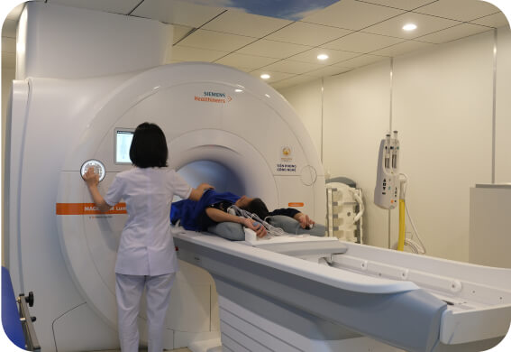
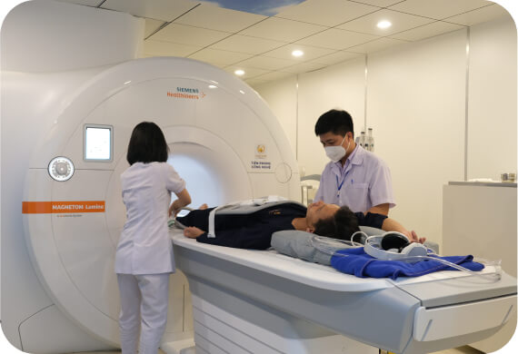
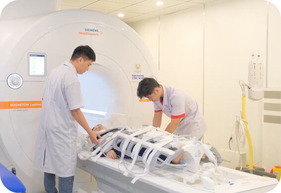
Images of the 3.0 Tesla Lumina MRI machine